The question
Does damaged alpha-synuclein protein cause Parkinson's nerve-cell loss?
This is the specific idea being tested.
Does damaged alpha-synuclein protein cause Parkinson's nerve-cell loss?
What you need to knowDamaged alpha-synuclein can matter, but Parkinson's appears to involve several routes to nerve-cell injury.Relative explanatory fit: Alternative ranked ahead
Why the alternative fits better: Protein build-up does not always match cell damage. Genes and cell vulnerability help explain that mismatch.
Claim assessed: Misfolded α-synuclein is directly neurotoxic to dopaminergic neurons, and its accumulation causes the neuronal loss of Parkinson's disease.
Finding: Scope-matched preregistration yields SC4 Γ for the full human direct-toxicity/PD-loss claim; SC1 Ζ for familial SNCA biology is preserved separately. The examined causal work establishes more than association, but does not yet isolate the specified endogenous human toxic mediator and survival dependency. Null trials are not treated as complete mechanistic refutations.
Evidence scope: 65 distinct primary reports actually examined: 25 focused partial full-text examinations and 40 abstracts/metadata; no full-paper/supplement audit claimed. Bounded primary-source search, not an exhaustive systematic review.
The grade applies to the stated claim. Model rankings and individual subclaim grades remain separate from the overall causal-evidence grade.
Full scientific assessment and references · Scorecard (JSON) · Review source (Markdown)
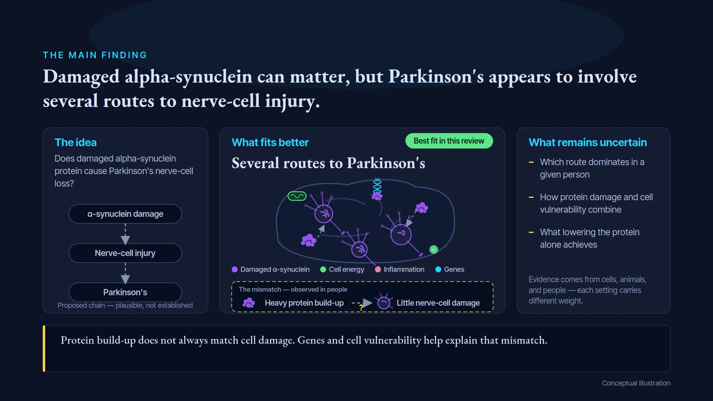
THE MAIN FINDING
Damaged alpha-synuclein can matter, but Parkinson's appears to involve several routes to nerve-cell injury.
The idea
Does damaged alpha-synuclein protein cause Parkinson's nerve-cell loss?
α-synuclein damage
Nerve-cell injury
Parkinson's
Proposed chain — plausible, not established
What fits better
Several routes to Parkinson's
Best fit in this review
Damaged α-synuclein
Cell energy
Inflammation
Genes
The mismatch — observed in people
Heavy protein build-up
Little nerve-cell damage
What remains uncertain
Which route dominates in a given person
How protein damage and cell vulnerability combine
What lowering the protein alone achieves
Evidence comes from cells, animals, and people — each setting carries different weight.
Protein build-up does not always match cell damage. Genes and cell vulnerability help explain that mismatch.
Conceptual illustration
This is the specific idea being tested.
Protein build-up does not always match cell damage. Genes and cell vulnerability help explain that mismatch.
A higher rank means a better fit to the evidence in this comparison. It does not prove every part of the leading explanation.
The reviewed hypothesis ranks 3 among 5 models compared; 2 alternative model(s) hold first rank for this stated claim.
Claim under review: rank 3 among 5 compared models.
Explanations sharing the lead: Heterogeneous PD pathways; Mitochondrial/lysosomal and proteostatic failure.
Position in this comparison: Next after leading rank.
The causal grade asks how well the stated causal claim is established. This comparison asks which of the assessed explanations fits the reviewed findings better. A leading explanation can remain unproven. Models can overlap or include the tested mechanism; an alternative ahead does not make that mechanism irrelevant. These ordinal ranks apply within this review; they do not measure the size of a difference or rank scientific strength across topics.
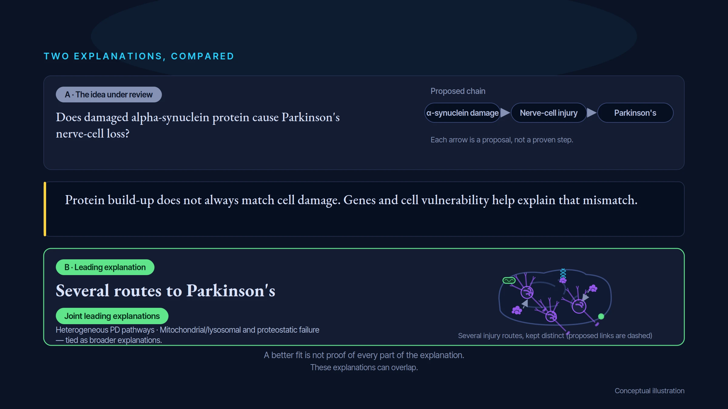
TWO EXPLANATIONS, COMPARED
A · The idea under review
Does damaged alpha-synuclein protein cause Parkinson's nerve-cell loss?
Proposed chain
α-synuclein damage
Nerve-cell injury
Parkinson's
Each arrow is a proposal, not a proven step.
Protein build-up does not always match cell damage. Genes and cell vulnerability help explain that mismatch.
B · Leading explanation
Several routes to Parkinson's
Joint leading explanations
Heterogeneous PD pathways · Mitochondrial/lysosomal and proteostatic failure — tied as broader explanations.
Several injury routes, kept distinct (proposed links are dashed)
A better fit is not proof of every part of the explanation.
These explanations can overlap.
Conceptual illustration
Plausible, not established for the stated claim.
Protein build-up does not always match cell damage. Genes and cell vulnerability help explain that mismatch.
These explanations can overlap. Ranking their fit to the evidence is separate from deciding how firmly a cause has been established.
Rank 1: Heterogeneous PD pathways — Distinct genetic, cellular and pathological routes converge on nigral vulnerability, with α-synuclein a context-dependent contributor.
Rank 1: Mitochondrial/lysosomal and proteostatic failure — Cellular dopamine/organellar/clearance failure contributes to injury and can promote α-synuclein accumulation as a mediator or response.
Rank 3: Direct misfolded α-synuclein toxicity — Misfolded α-synuclein is directly neurotoxic to dopaminergic neurons, and its accumulation causes the neuronal loss of Parkinson's disease.
Rank 4: Inclusion sequestration can protect — Some aggregation or inclusion formation sequesters harmful soluble protein/material, so total visible burden need not equal toxic exposure.
Rank 5: Glial/non-cell-autonomous toxicity — Host inflammatory or glial responses contribute to injury rather than all loss arising solely through direct neuron-intrinsic toxicity.
Evidence rating: Γ · Moderate confidence. How the evidence was assessed.
Protein build-up does not always match cell damage. Genes and cell vulnerability help explain that mismatch.
This is an assessment of a causal explanation, not proof that a particular supplement, diet, test, or treatment improves health. A treatment claim needs studies that test the intervention in people and measure meaningful benefits and harms. These findings can help frame research and informed clinical questions, but they do not identify what is right for one person.
Look for the proposed factor and the outcome, in the people or systems covered by the claim.
Several routes to Parkinson's. Protein build-up does not always match cell damage. Genes and cell vulnerability help explain that mismatch.
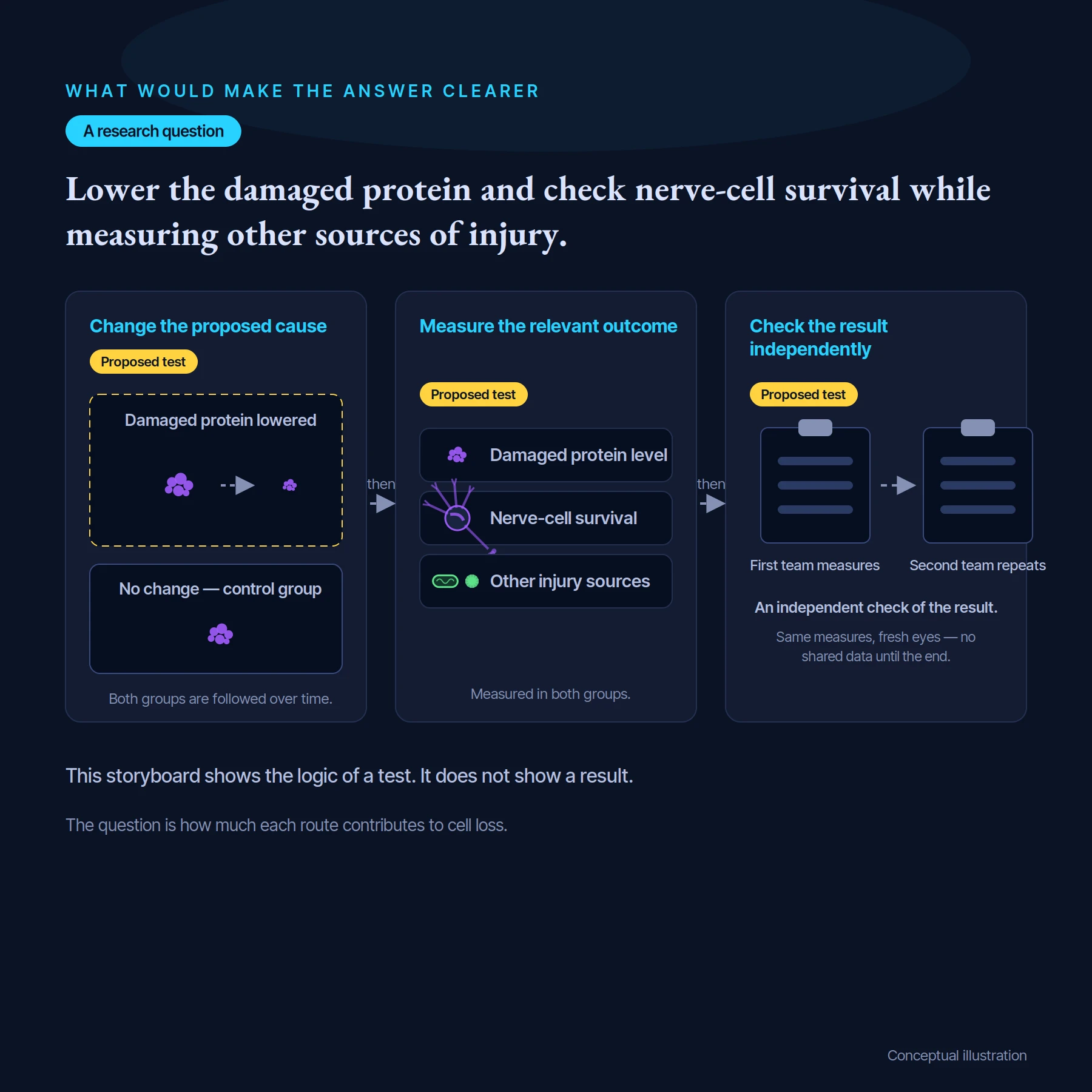
Lower the damaged protein and check nerve-cell survival while measuring other sources of injury.
These illustrations show the key questions tested in the review. They are conceptual drawings, not microscope images or experimental results.
Endogenous changes in SNCA dosage or sequence cause a dopaminergic degenerative disease phenotype through α-synuclein biology.
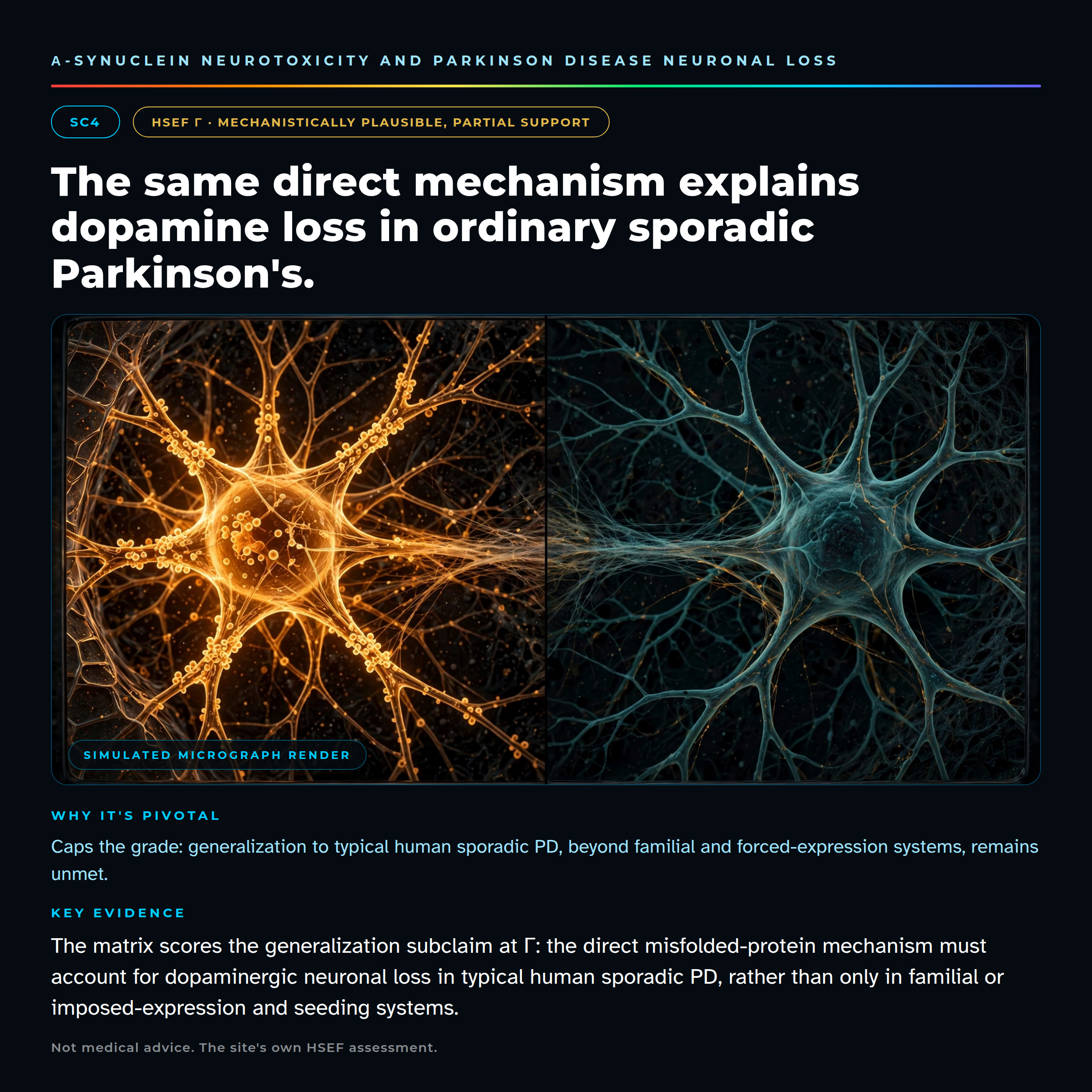
The direct misfolded-protein mechanism accounts for dopaminergic neuronal loss in typical human sporadic PD, rather than only familial or imposed-expression/seeding systems.
WHAT WOULD MAKE THE ANSWER CLEARER
A research question
Lower the damaged protein and check nerve-cell survival while measuring other sources of injury.
then
then
Change the proposed cause
Proposed test
Damaged protein lowered
No change — control group
Both groups are followed over time.
Measure the relevant outcome
Damaged protein level
Nerve-cell survival
Other injury sources
Measured in both groups.
Check the result independently
Proposed test
First team measures
Second team repeats
An independent check of the result.
Same measures, fresh eyes — no shared data until the end.
This storyboard shows the logic of a test. It does not show a result.
The question is how much each route contributes to cell loss.
Conceptual illustration
Lower the damaged protein and check nerve-cell survival while measuring other sources of injury.
Check that the intended change really happened.
Allow for other factors and compare with suitable controls.
Look for independent confirmation and the limits of the effect.
These are questions for research, not a claim that the proposed studies have already succeeded.
Proposed tests: 2 test design(s) are registered in the scorecard.
In plain terms, researchers would change the proposed cause, check that it changed as intended, then measure a meaningful outcome while controlling plausible alternatives. The result should be able to support or count against the claim—not only show that a pathway exists.
Study design: In a physiological SNCA-expression PD-relevant system, selectively remove a molecularly defined harmful conformer while preserving monomeric synaptic function, then restore only that conformer without raising total protein or causing nonspecific proteostatic injury.
What would be measured: Orthogonal conformer abundance/occupancy, longitudinal dopaminergic neuron lineage survival, pan-neuronal counts, dopamine physiology and sustained function.
Important controls: Toxicity and efficacy independent of TH expression; equal soluble protein/function controls, inactive reagent, matched conformer versus nontoxic species rescue and blinded exposure/timing.
Result that would support the claim: Selective removal preserves neurons and matched harmful-conformer reinstatement restores loss, with a nontoxic species failing to do so.
Result that would count against it: Effective specific conformer removal leaves independently measured neuron loss unchanged, and matched reinstatement does not restore injury.
Study design: Preregister baseline PD/genotype/seed strata and an adequate clinical horizon; randomize an intervention with validated engagement of the proposed intracellular/extracellular toxic species and an orthogonal normal-function-preserving strategy.
What would be measured: Primary disease trajectory, a validated degeneration endpoint and pathogenic-species engagement measured separately; causal mediation addressing symptoms, inflammation and total-protein effects.
Important controls: Do not define responders after treatment; free CSF signal or seeding fluorescence is not automatically quantitative brain conformer removal; independently validate the degeneration endpoint.
Result that would support the claim: A replicated primary trajectory/degeneration benefit explained by the selective toxic mediator rather than symptomatic or general anti-inflammatory action.
Result that would count against it: Verified adequate harmful-species depletion without clinical/degeneration improvement under prespecified exposure and stage.
Follow the studies, detailed comparisons and evidence ratings behind this guide.
Open the scientific review →