The question
Does loosening tightly packed DNA drive normal ageing?
This is the specific idea being tested.
Does loosening tightly packed DNA drive normal ageing?
What you need to knowDNA packaging changes may add to ageing-related harm, but are not established as the main ageing clock.Relative explanatory fit: Alternative ranked ahead
Why the alternative fits better: Loss of gene silencing can release normally quiet DNA activity and amplify inflammation.
Claim assessed: Progressive loss of H3K9me3, H3K27me3 and HP1-marked repressive heterochromatin is a primary causal driver of organismal chronological aging.
Finding: The registered scope-matched gate is natural chronological primacy SC3, assessed Γ. Native old MuSC mediation supports a real scoped contribution and adult Dicer-2 supports lifespan intervention, but neither isolates gradual global mark erosion as the primary organismal clock. Calibrated H3K27me3 gain, mark redistribution and null longevity contrasts defeat the universal net-loss premise. Adult global triple-knockout evidence is a preprint outside the 60 peer-reviewed count; the index empirical anchor cannot silently be upgraded.
Evidence scope: 60 primary reports; literature cutoff 2026-10-09; source examination UTC date 2026-10-09; bounded critical review, not exhaustive systematic review.
The grade applies to the stated claim. Model rankings and individual subclaim grades remain separate from the overall causal-evidence grade.
Full scientific assessment and references · Scorecard (JSON) · Review source (Markdown)
THE MAIN FINDING
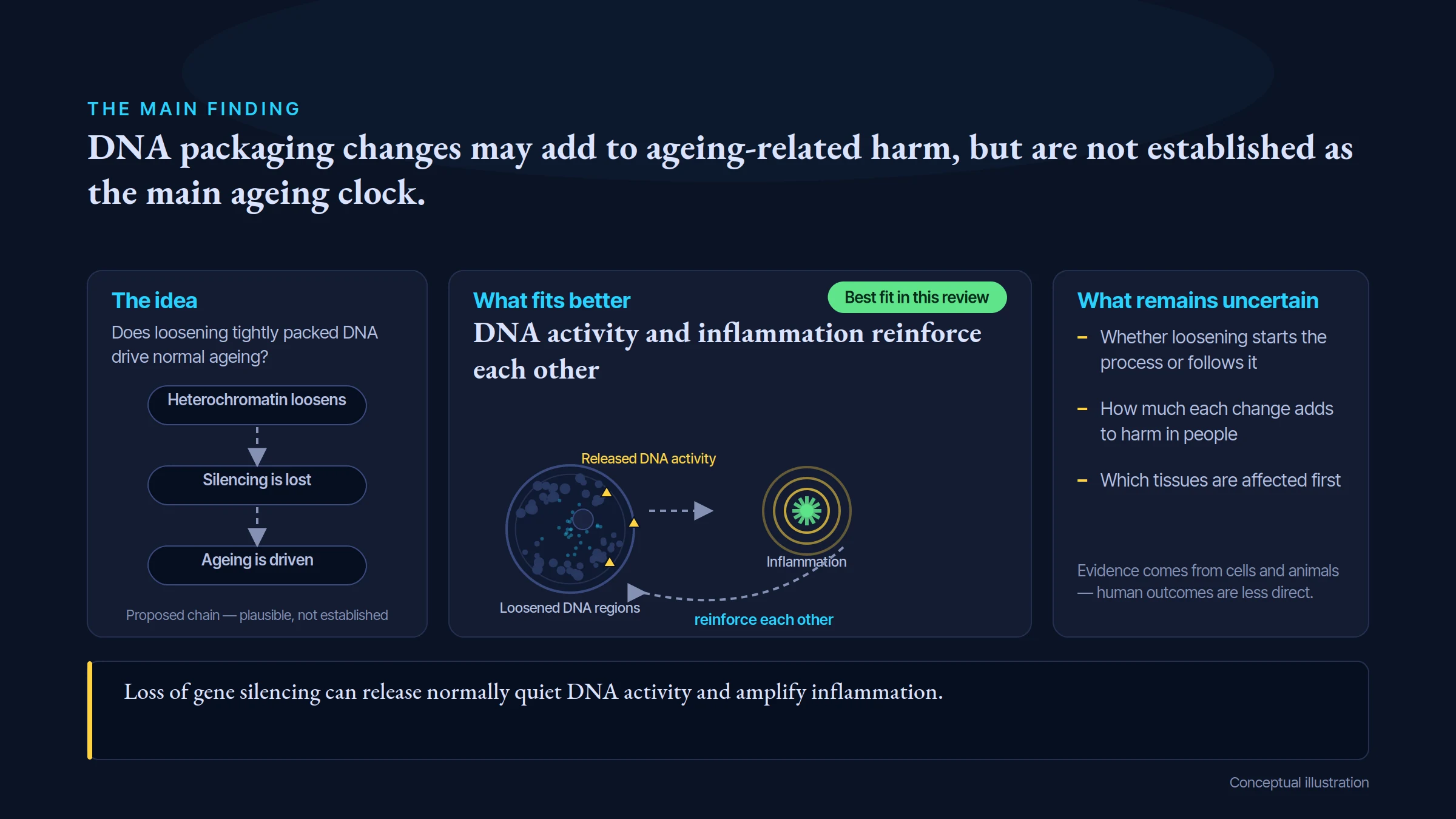
DNA packaging changes may add to ageing-related harm, but are not established as the main ageing clock.
The idea
What fits better
What remains uncertain
Does loosening tightly packed DNA drive normal ageing?
Heterochromatin loosens
Silencing is lost
Ageing is driven
Proposed chain — plausible, not established
DNA activity and inflammation reinforce each other
Best fit in this review
Loosened DNA regions
Released DNA activity
Inflammation
reinforce each other
Whether loosening starts the process or follows it
How much each change adds to harm in people
Which tissues are affected first
Evidence comes from cells and animals — human outcomes are less direct.
Loss of gene silencing can release normally quiet DNA activity and amplify inflammation.
Conceptual illustration
This is the specific idea being tested.
Loss of gene silencing can release normally quiet DNA activity and amplify inflammation.
A higher rank means a better fit to the evidence in this comparison. It does not prove every part of the leading explanation.
The reviewed hypothesis ranks 5 among 5 models compared; 1 alternative model(s) hold first rank for this stated claim.
Claim under review: rank 5 among 5 compared models.
Leading explanation: TE/innate-immune feedback.
Position in this comparison: Last recorded rank.
The causal grade asks how well the stated causal claim is established. This comparison asks which of the assessed explanations fits the reviewed findings better. A leading explanation can remain unproven. Models can overlap or include the tested mechanism; an alternative ahead does not make that mechanism irrelevant. These ordinal ranks apply within this review; they do not measure the size of a difference or rank scientific strength across topics.
TWO EXPLANATIONS, COMPARED
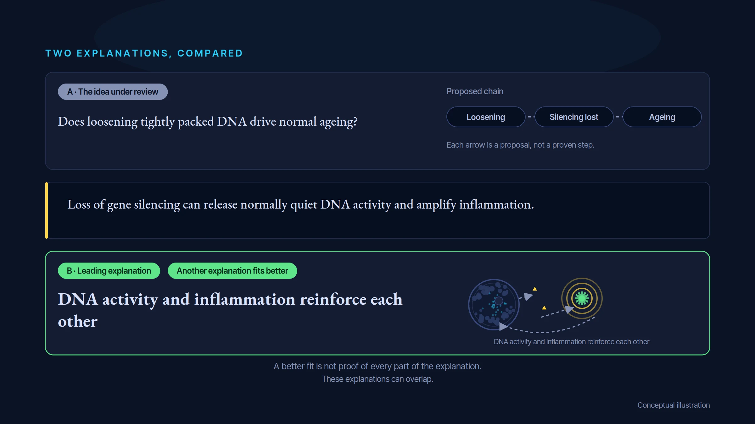
A · The idea under review
Does loosening tightly packed DNA drive normal ageing?
Proposed chain
Loosening
Silencing lost
Ageing
Each arrow is a proposal, not a proven step.
Loss of gene silencing can release normally quiet DNA activity and amplify inflammation.
B · Leading explanation
DNA activity and inflammation reinforce each other
Another explanation fits better
DNA activity and inflammation reinforce each other
A better fit is not proof of every part of the explanation.
These explanations can overlap.
Conceptual illustration
Plausible, not established for the stated claim.
Loss of gene silencing can release normally quiet DNA activity and amplify inflammation.
These explanations can overlap. Ranking their fit to the evidence is separate from deciding how firmly a cause has been established.
Rank 1: TE/innate-immune feedback — Loss of repression and TE products amplify senescent/inflammatory states.
Rank 2: Mark redistribution and tissue-dependent remodeling — Aging alters location and identity of repressive chromatin rather than uniformly erasing it.
Rank 3: Metabolic and DNA-damage triage — Metabolism/stress drives epigenomic changes which can feed back on function.
Rank 4: Lamina/LAD repositioning — Nuclear tethering affects repression and inflammatory outputs.
Rank 5: Global erosion primary clock — Progressive global repressive-mark loss drives chronological organismal aging.
Evidence rating: Γ · Moderate confidence. How the evidence was assessed.
Loss of gene silencing can release normally quiet DNA activity and amplify inflammation.
The registered scope-matched gate is natural chronological primacy SC3, assessed Γ. Native old MuSC mediation supports a real scoped contribution and adult Dicer-2 supports lifespan intervention, but neither isolates gradual global mark erosion as the primary organismal clock. Calibrated H3K27me3 gain, mark redistribution and null longevity contrasts defeat the universal net-loss premise. Adult global triple-knockout evidence is a preprint outside the60 peer-reviewed count; the index empirical anchor cannot silently be upgraded.
Look for the proposed factor and the outcome, in the people or systems covered by the claim.
DNA activity and inflammation reinforce each other. Loss of gene silencing can release normally quiet DNA activity and amplify inflammation.
Restore a defined part of DNA silencing and measure function without changing other repair processes.
WHAT WOULD MAKE THE ANSWER CLEARER
A research question
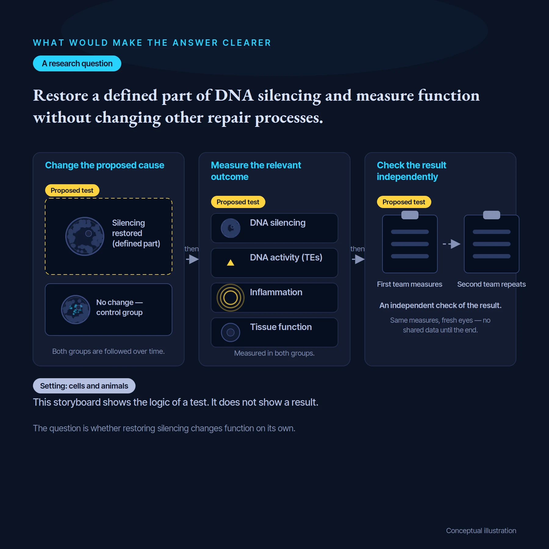
Restore a defined part of DNA silencing and measure function without changing other repair processes.
then
then
Change the proposed cause
Proposed test
Measure the relevant outcome
Proposed test
Check the result independently
Proposed test
Silencing restored (defined part)
No change — control group
DNA silencing
DNA activity (TEs)
Inflammation
Tissue function
Measured in both groups.
First team measures
Second team repeats
An independent check of the result.
Same measures, fresh eyes — no shared data until the end.
Both groups are followed over time.
Setting: cells and animals
This storyboard shows the logic of a test. It does not show a result.
The question is whether restoring silencing changes function on its own.
Conceptual illustration
Restore a defined part of DNA silencing and measure function without changing other repair processes.
Check that the intended change really happened.
Allow for other factors and compare with suitable controls.
Look for independent confirmation and the limits of the effect.
These are questions for research, not a claim that the proposed studies have already succeeded.
Proposed tests: 1 test design(s) are registered in the scorecard.
In plain terms, researchers would change the proposed cause, check that it changed as intended, then measure a meaningful outcome while controlling plausible alternatives. The result should be able to support or count against the claim—not only show that a pathway exists.
Study design: Register normal adult mouse cohorts of both sexes and prespecified proliferative/postmitotic tissues. Restore H3K9 repression at native levels selectively, cross with TE blockade and SAM normalization, and use orthogonal mark-selective rescue without changing growth or caloric intake.
What would be measured: Calibrated multi-mark quantity and distribution, TE RNA/cDNA, innate activation, lifespan, frailty and uninjured tissue function
Important controls: Adult timing, littermate/background matching, expression/binding-matched inactive rescue, equal intake, cell composition, injury-free functional assessment and dose gradient
Result that would support the claim: Selective preservation slows prespecified natural trajectories and orthogonal rescue restores function beyond TE/metabolic effects across tissues
Result that would count against it: Marks change without function, benefits require injury/developmental growth change, or TE/metabolic rescue works despite unrepaired chromatin
Follow the studies, detailed comparisons and evidence ratings behind this guide.
Open the scientific review →