The question
Does healthy brain tissue contain resident microbes that drive degeneration?
This is the specific idea being tested.
Does healthy brain tissue contain resident microbes that drive degeneration?
What you need to knowDetecting microbial material does not establish a living microbial community in healthy brain tissue.Relative explanatory fit: Alternative ranked ahead
Why the alternative fits better: Reagents, handling and host material can create apparent microbial profiles when the true signal is very small.
Claim assessed: The living human brain harbors a resident microbial community that contributes causally to neurodegeneration.
Finding: Preregistered primary-subclaim aggregation returns Α because SC1 living-human community existence remains unresolved under the low-biomass contamination gate. SC2 Β and SC3 Γ do not replace that prerequisite. Weakest-link and scope-matched sensitivities also return Α. Positive fish ecology and AD autopsy culture are explicitly retained rather than dismissed.
Evidence scope: 59 primary reports; literature cutoff 2026-10-08; source examination UTC date 2026-10-08; bounded critical review, not exhaustive systematic review.
The grade applies to the stated claim. Model rankings and individual subclaim grades remain separate from the overall causal-evidence grade.
Full scientific assessment and references · Scorecard (JSON) · Review source (Markdown)
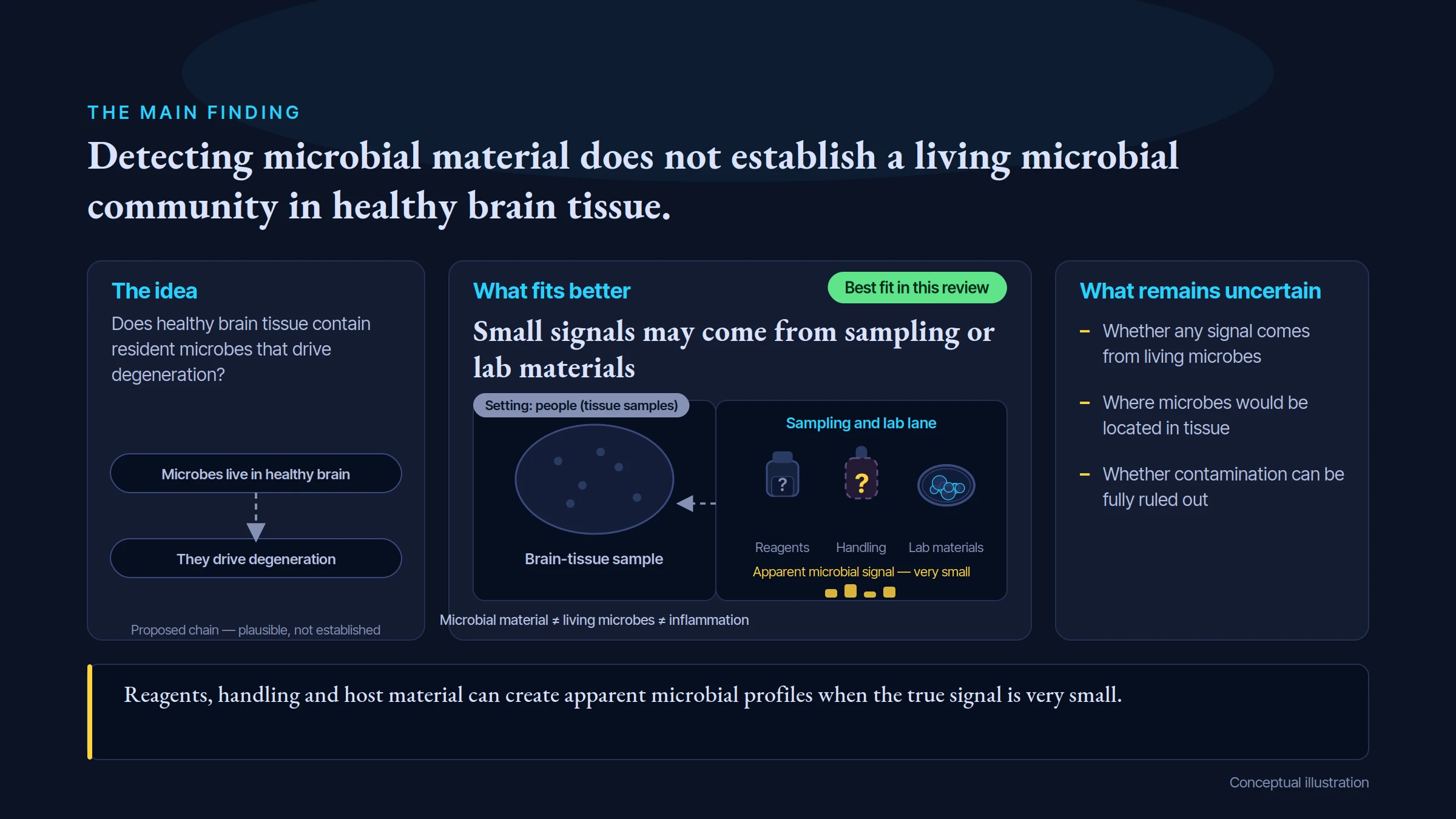
THE MAIN FINDING
Detecting microbial material does not establish a living microbial community in healthy brain tissue.
The idea
Does healthy brain tissue contain resident microbes that drive degeneration?
Microbes live in healthy brain
They drive degeneration
Proposed chain — plausible, not established
What fits better
Small signals may come from sampling or lab materials
Best fit in this review
Brain-tissue sample
Sampling and lab lane
Reagents
Handling
Lab materials
Apparent microbial signal — very small
Microbial material, living microbes and inflammation are distinct
Setting: people (tissue samples)
What remains uncertain
Whether any signal comes from living microbes
Where microbes would be located in tissue
Whether contamination can be fully ruled out
Reagents, handling and host material can create apparent microbial profiles when the true signal is very small.
Conceptual illustration
This is the specific idea being tested.
Reagents, handling and host material can create apparent microbial profiles when the true signal is very small.
A higher rank means a better fit to the evidence in this comparison. It does not prove every part of the leading explanation.
The reviewed hypothesis ranks 5 among 5 models compared; 1 alternative model(s) hold first rank for this stated claim.
Claim under review: rank 5 among 5 compared models.
Leading explanation: Low-biomass reagent/host artifact.
Position in this comparison: Last recorded rank.
The causal grade asks how well the stated causal claim is established. This comparison asks which of the assessed explanations fits the reviewed findings better. A leading explanation can remain unproven. Models can overlap or include the tested mechanism; an alternative ahead does not make that mechanism irrelevant. These ordinal ranks apply within this review; they do not measure the size of a difference or rank scientific strength across topics.
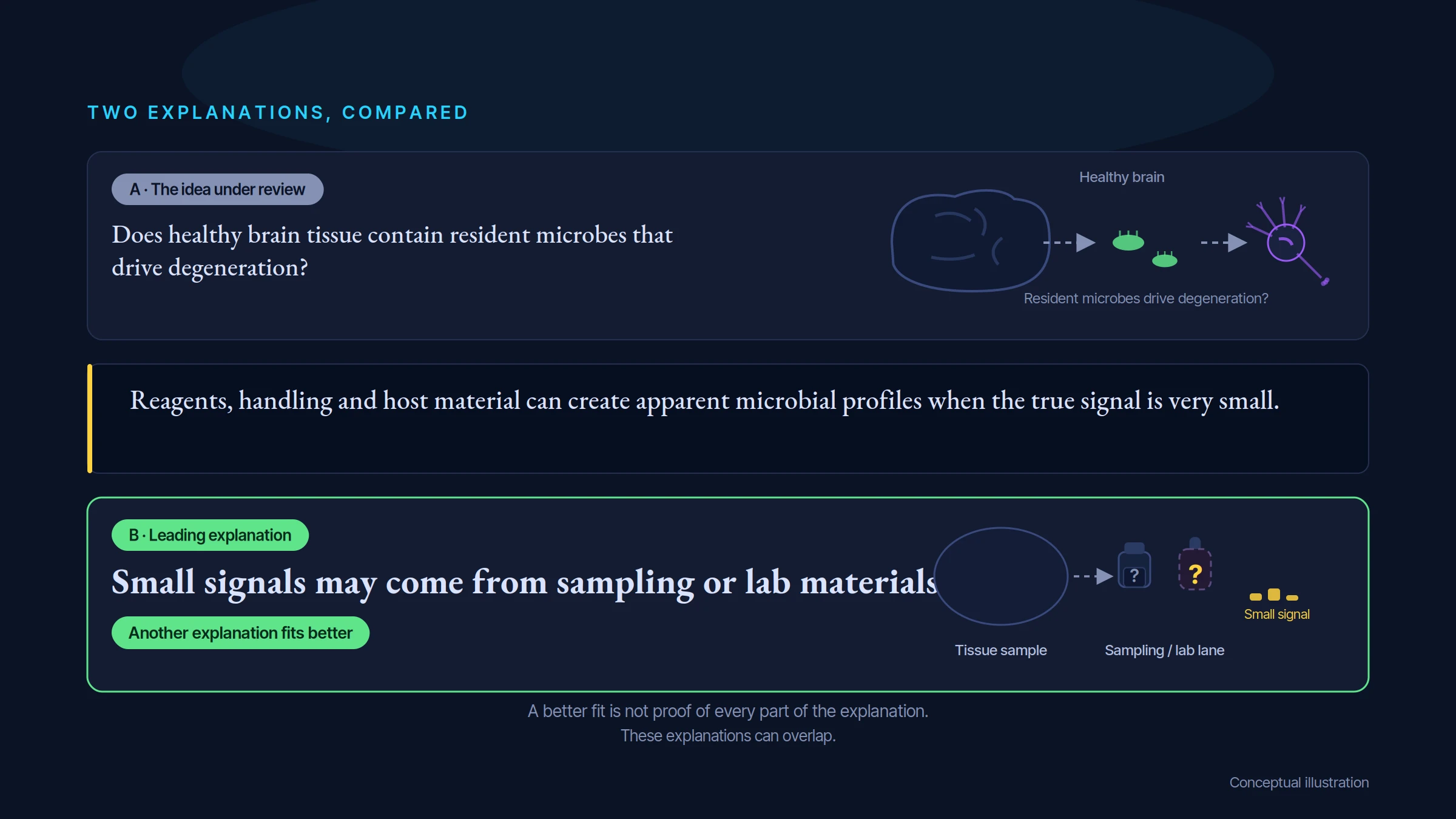
TWO EXPLANATIONS, COMPARED
A · The idea under review
Does healthy brain tissue contain resident microbes that drive degeneration?
Healthy brain
Resident microbes drive degeneration? (proposed, not established)
Reagents, handling and host material can create apparent microbial profiles when the true signal is very small.
B · Leading explanation
Small signals may come from sampling or lab materials
Another explanation fits better
Tissue sample
Sampling and lab materials
Small signal may come from sampling or lab materials
A better fit is not proof of every part of the explanation.
These explanations can overlap.
Conceptual illustration
Not established for the stated claim.
Reagents, handling and host material can create apparent microbial profiles when the true signal is very small.
These explanations can overlap. Ranking their fit to the evidence is separate from deciding how firmly a cause has been established.
Rank 1: Low-biomass reagent/host artifact — Several technical sources can generate reproducible apparent bacterial profiles.
Rank 2: Disease-related transient microbial ingress — Pathogens or their molecules reach brain in selected disease settings without a stable community.
Rank 3: Teleost-restricted residency — Healthy salmonid brain can contain viable bacteria under the reported conditions.
Rank 4: Agonal/postmortem translocation — Cadaver communities and handling change tissue microbial signals.
Rank 5: Resident human brain community — A viable stable brain community contributes to neurodegeneration.
Evidence rating: Α · Moderate confidence. How the evidence was assessed.
Reagents, handling and host material can create apparent microbial profiles when the true signal is very small.
A result about how a disease works is a separate question from whether a treatment improves health.
Look for the proposed factor and the outcome, in the people or systems covered by the claim.
Small signals may come from sampling or lab materials. Reagents, handling and host material can create apparent microbial profiles when the true signal is very small.
Use independent laboratories and sampling controls, showing living microbes at the same tissue location.
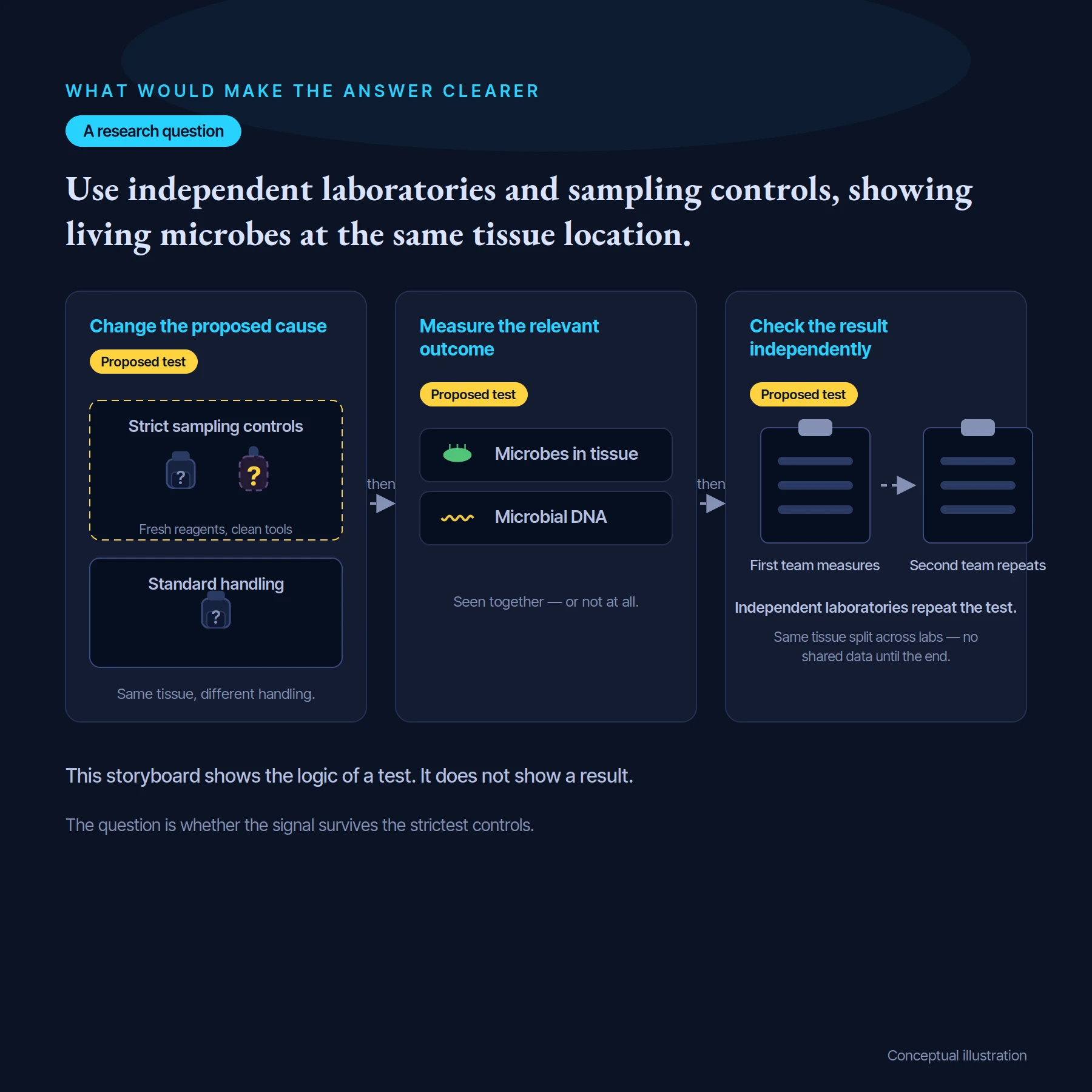
WHAT WOULD MAKE THE ANSWER CLEARER
A research question
Use independent laboratories and sampling controls, showing living microbes at the same tissue location.
then
then
Change the proposed cause
Proposed test
Strict sampling controls
Fresh reagents, clean tools
Standard handling
Same tissue, different handling.
Measure the relevant outcome
Proposed test
Microbes in tissue
Microbial DNA
Seen together — or not at all.
Check the result independently
Proposed test
First team measures
Second team repeats
Independent laboratories repeat the test.
Same tissue split across labs — no shared data until the end.
This storyboard shows the logic of a test. It does not show a result.
The question is whether the signal survives the strictest controls.
Conceptual illustration
Use independent laboratories and sampling controls, showing living microbes at the same tissue location.
Check that the intended change really happened.
Allow for other factors and compare with suitable controls.
Look for independent confirmation and the limits of the effect.
These are questions for research, not a claim that the proposed studies have already succeeded.
Proposed tests: 1 test design(s) are registered in the scorecard.
In plain terms, researchers would change the proposed cause, check that it changed as intended, then measure a meaningful outcome while controlling plausible alternatives. The result should be able to support or count against the claim—not only show that a pathway exists.
Study design: A prospectively registered multi-laboratory living-tissue study coupling quantitative strain-resolved genomes/RNA, recoverable viability and in situ localization to matched controls, followed by selective removal/reconstitution in a biologically matched model only after existence is demonstrated.
What would be measured: Absolute organism recovery above validated protocol background, viable strain concordance, vascular exclusion and separate neuronal/functional endpoints
Important controls: Independent collection/tool/reagent/PCR blanks, host-aware mapping, blind spike-ins with difficult-to-lyse taxa, matched culture controls and same-donor heat comparisons
Result that would support the claim: The same viable localized strains reproducibly exceed controls across laboratories and selective removal/rescue moves neurodegenerative phenotype
Result that would count against it: Signal follows lots, host sequences, blood or collection delay, or confirmed removal leaves disease intact
Follow the studies, detailed comparisons and evidence ratings behind this guide.
Open the scientific review →